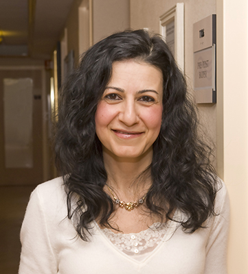
 Edna Kapenhas, MD, leads the breast surgery and breast surgical oncology program at Stony Brook Southampton Hospital. She is also the medical director of The Ellen Hermanson Breast Center. She attended Queens College and earned her medical degree from State University at Buffalo. She completed her surgical residency training at New York Hospital of Queens, followed by a yearlong clinical fellowship in breast surgery at St. Luke’s-Roosevelt Hospital Center and Beth Israel Medical Center. One of her many areas of interest is prevention and early detection of breast cancer. She is actively involved in clinical research.
Dr. Kapenhas sees patients in Southampton and Riverhead. To make an appointment, please call (631) 726-8400.
What new technologies have been introduced at The Ellen Hermanson Breast Center over the last five years?
One of the new things is tomosynthesis, which is a state-of-the art mammogram that gives a 3D image of the breast. It is superior to other forms of breast imaging. Tomosynthesis is available at The Ellen Hermanson Breast Center at the Hospital, the Medical Atrium in Hampton Bays, and in East Hampton. Another exciting technology —it’s less than a year old—is our stereotactic biopsy system, located at the Hospital, which works with the 3D mammography to precisely identify and biopsy any abnormalities in the breast.
In the past, sonography could differentiate a cystic mass versus a solid one. In case of a solid mass, you had to decide if you would proceed with a biopsy or a 6-month follow-up imaging appointment. Now we have a state-of-the-art sonography machine designed by a French engineer especially for breast imaging. One of the features is elastography, which can detect the elasticity of the tissue surrounding a mass and categorize whether the lesion is likely benign or suspicious. A suspicious lesion—for example, cancer—can actually infiltrate into the surrounding tissue and make it more rigid, which can be identified by elastography.
For more precision in localization of non-palpable lesions in the breast prior to surgical procedures, we have been using a radioactive seed, which is basically a little seed that has low-grade radioactivity. It is inserted into the lesion by a radiologist, and I have a handheld Geiger counter that picks up the radioactivity localizing the exact location. It’s great for the patient because it saves time and is more comfortable. If a patient has needle localization, it has to be inserted in the morning, so it delays surgical time, and it’s also uncomfortable for the patient to sit and wait for surgery with a needle in her breast. The needle can also become dislodged. The radioactive seed can be inserted a day or two before the surgery and is a very accurate localization of the lesion. Currently we’re also trying another technology, an infrared activated reflector, that works on radar technology. It’s nonradioactive and inserted by a radiologist a well.
When and how often should a woman get a mammography?
For women who are asymptomatic and have no family history, I would recommend a mammography starting at age 40 and every year thereafter. For people who have family history, the recommendation is to start 10 years younger than the age of diagnosis for first-degree relatives. For example, if a mother was diagnosed at age 40, we would start screening the daughters at age 30. It’s important because early detection is key—the earlier breast cancer is detected, the higher the chance of being able to save the breast, as well as the higher chance of survival. One year is enough for a cancer to develop—that’s why we recommend a mammography once a year. If you get your mammography every year, and you develop cancer in between screenings, it is more likely to be in an early stage.
With the new tomosynthesis machine we have now, the process is quicker. The pedals that are used to compress the breast are more comfortable. They’re still squeezing the breast, but it’s not as uncomfortable as before. The breast needs to be compressed, so the tissue is spread, out and the mammography can visualize if there’s anything within the breast tissue. In general, it’s very quick—before you know it, it’s over. Mammography is much better tolerated nowadays with 3D than before.
Does having breast implants affect the readability of a mammogram?
Yes, it can. Having breast implants can make it harder because the implant can be in the way—you have to get additional images and displaced views to push the implant out of the way to visualize the breast tissue. This maybe more pronounced in some cases depending on the location of the implant, particularly if it’s behind or in front of the muscle. If it is an old implant and it is encapsulated and hard, it makes the process of imaging even more difficult. Mammography is still the gold standard. If a woman with breast implants gets an MRI, she still needs to undergo her yearly mammography, as certain findings can be detected on a mammogram and not a MRI, and vice versa.
Do you recommend self-breast exams between screenings?
I advise patients to do a self-breast exam once a month. If from one month to the next you feel something different, please make an appointment and let’s find out what it is. Hopefully it will be nothing, but if it does require attention, early treatment results in better outcomes.
When I do a breast exam on my patients, I do it multi-positional: I examine them sitting up and laying down with their arm up. When they do it on their own, they can look in the mirror, put their arms up, or put their hands on their hips to see if there is any dimpling in the skin, any redness or skin thickening, or nipple retraction or inversion. And then when they feel, they want to not only feel for lumps, but they want to see if any area feels firm or hard. Is there any nipple discharge? (Squeezing the nipple is not recommended.) Bloody nipple discharge is alarming. Be sure and check the lymph nodes in the armpits. Use the pads of your three longest fingers and apply moderate pressure.
How will The Ellen Hermanson Foundation be involved with the new Phillips Family Cancer Center?
Treating breast cancer is a multi-disciplinary team approach involving a breast surgeon, a medical and radiation oncologist, and many other services. The Phillips Family Cancer Center will have chemotherapy infusion suites, as well as a radiation center. Although most patients diagnosed with breast cancer undergo surgery prior to any other treatment, some patients require chemotherapy before surgery. In most cases of early stage breast cancer, breast conservation can be performed. In this procedure the cancer is surgically removed with a small amount of surrounding normal breast tissue and the rest of the breast is saved. These patients will then require radiation therapy. Some women after breast cancer surgery will need both chemotherapy and radiation, and others just radiation. The majority of breast cancers are hormone receptor positive, so these patients will also require anti-hormonal therapy—which is in the form of a pill taken orally—for at least five years. Once The Phillips Family Cancer Center opens, a woman will be able to get all of this treatment without leaving the South Fork.
Chemotherapy and radiation treatments can take a toll on a woman’s body physically. But how does breast cancer affect a woman emotionally?
Most women look at their breasts as a sign of their femininity. When talking to someone who is BRCA positive—with the breast cancer gene—about having a bilateral mastectomy and an oophorectomy (the removal of ovaries is recommended because the patient who carries the gene is at high risk for ovarian cancer, in addition to breast cancer), it’s easier for some patients to accept the concept of losing their ovaries, but not their breasts. Losing their hair is another emotional loss tied to self-identity. When a woman learns she has cancer, she immediately thinks she may lose one or both breasts and her hair. It’s very, very emotionally draining. For the mastectomy patients, I tell them reconstruction is an option. I think having that option helps them deal with the situation better. As for losing their hair, I suggest patients consider cutting it short before chemo, but to keep in mind it will grow back when they finish their treatment. And it actually comes back better!
Everyone seems to know someone who has been diagnosed with breast cancer. So why is it important to continue raising awareness?
One in 8 U.S. women will develop invasive breast cancer over the course of her lifetime. It is important to keep raising awareness so people keep getting their mammograms and are aware that if they feel something in their breasts, they need to seek immediate medical attention. That helps us get better at picking it up earlier and earlier, translating to a higher cure rate and decreased mortality. Breast cancer mortality rates have decreased over the years, and that’s thought to be the result of not only treatment advances, but earlier detection through screening and increased awareness.

